
Study Illuminates the Structural Features of Memory Formation at the Cellular and Subcellular Levels
NIH-funded study uses cutting-edge imaging techniques to reconstruct features underlying learning and memory in the mouse brain
• Media Advisory
What:
In a study supported by the National Institutes of Health (NIH), researchers revealed the structural underpinnings of memory formation across a broad network of neurons in the mouse brain. This work sheds light on the fundamentally flexible nature of how memories are made, detailing learning-related changes at the cellular and subcellular levels with unprecedented resolution. Understanding this flexibility may help explain why memory and learning processes sometimes go awry.
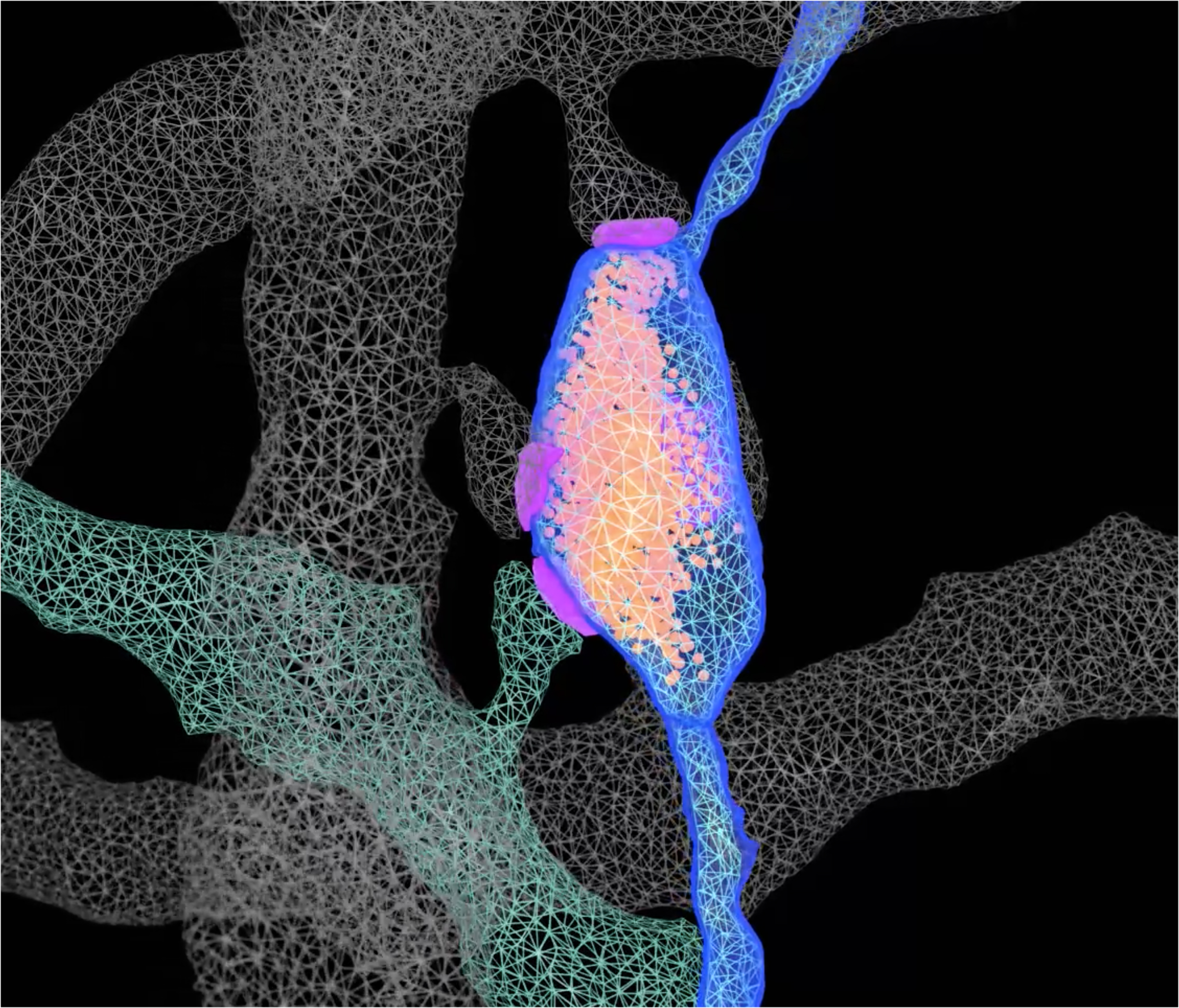
The findings, published in Science, showed that neurons assigned to a memory trace reorganized their connections to other neurons through an atypical type of connection called a multi-synaptic bouton. In a multi-synaptic bouton, the axon of the neuron relaying the signal with information contacts multiple neurons that receive the signal. According to the researchers, multi-synaptic boutons may enable the cellular flexibility of information coding observed in previous research.
The researchers also found that neurons involved in memory formation were not preferentially connected with each other. This finding challenges the idea that “neurons that fire together wire together,” as would be predicted by a traditional theory of learning.
In addition, the researchers observed that neurons allocated to a memory trace reorganized certain intracellular structures that provide energy and support communication and plasticity in neuronal connections. These neurons also had enhanced interactions with support cells known as astrocytes.
Using a combination of advanced genetic tools, 3D electron microscopy, and artificial intelligence, Scripps Research scientists Marco Uytiepo, Anton Maximov, Ph.D., and colleagues reconstructed a wiring diagram of neurons involved in learning and identified structural changes to these neurons and their connections at the cellular and subcellular levels.
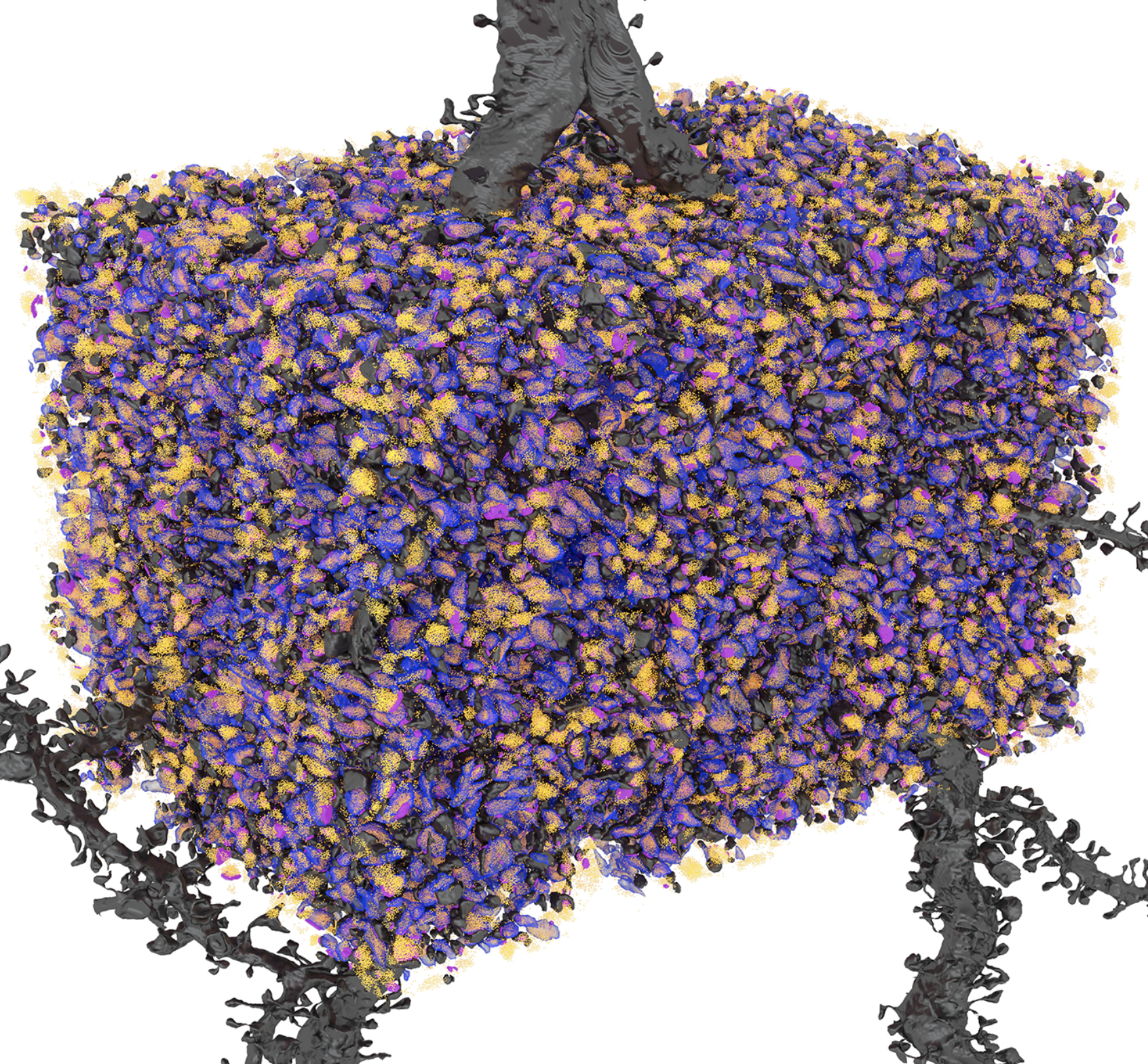
To examine structural features associated with learning, the researchers exposed mice to a conditioning task and examined the hippocampus region of the brain about 1 week later. They selected this time point because it occurs after memories are first encoded but before they are reorganized for long-term storage. Using advanced genetic techniques, the researchers permanently labeled subsets of hippocampal neurons activated during learning, which enabled reliable identification. They then used 3D electron microscopy and artificial intelligence algorithms to produce nanoscale reconstructions of the excitatory neural networks involved in learning.
This study provides a comprehensive view of the structural hallmarks of memory formation in one brain region. It also raises new questions for further exploration. Future studies will be crucial in determining whether similar mechanisms operate across different time points and neural circuits. In addition, further investigation into the molecular composition of multi-synaptic boutons is needed to determine their precise role in memory and other cognitive processes.
The research was supported by funding from the National Institute of Mental Health, the National Institute of Neurological Disorders and Stroke, and NIH’s Brain Research Through Advancing Innovative Neurotechnologies® Initiative, or The BRAIN Initiative®.
Who:
Jamie Driscoll, National Institute of Mental Health
Dr. Eunyoung Kim, National Institute of Mental Health
Study:
Uytiepo, M., Zhu, Y., Bushong, E., Chou, K., Polli, F. S., Zhao, E., Kim, K.-Y., Luu, D., Chang, L., Yang, D., Ma, T. C., Kim, M., Zhang, Y., Walton, G., Quach, T., Haber, M., Patapoutian, L., Shahbazi, A., Zhang, Y., … Maximov, A. (2025). Synaptic architecture of a memory engram in the mouse hippocampus. Science. http://www.science.org/doi/10.1126/science.ado8316
NIH funding:
###
The Brain Research Through Advancing Innovative Neurotechnologies® and The BRAIN Initiative® are registered trademarks of HHS.
About the National Institute of Mental Health (NIMH): The mission of the NIMH is to transform the understanding and treatment of mental illnesses through basic and clinical research, paving the way for prevention, recovery and cure. For more information, visit the NIMH website.
About the National Institutes of Health (NIH): NIH, the nation's medical research agency, includes 27 Institutes and Centers and is a component of the U.S. Department of Health and Human Services. NIH is the primary federal agency conducting and supporting basic, clinical, and translational medical research, and is investigating the causes, treatments, and cures for both common and rare diseases. For more information about NIHand its programs, visit the NIH website .
NIH…Turning Discovery Into Health®