
Equipment and Image Processing
Image Acquisition Capabilities
- High-throughput automated slide scanning. Up to 100 slides can be loaded at one time. Epifluorescence with excitation from 405 through 730nm and brightfield are available. Stitching, extended depth of field projection (EDF), and channel unmixing are available.
- Confocal microscopy. Instruments capable of precise excitation and emission collection in conjunction with resonant scanning, optical sectioning, time gating, fluorescence lifetime imaging microscopy (FLIM), deep tissue, and whole mouse organ imaging are available.
- Single-plane illumination (Lightsheet) microscopy optimized for imaging large cleared tissue, including whole mouse brains and substantial parts of brains from other species is availalbe.
Image Analysis Resources
- Use of dedicated image processing and analysis computer workstations is available remotely or in-person.
- Several commercial image processing and analysis software packages are available, with features that include automatic cell detection, semiautomated neuronal morphology tracing, and rodent brain atlas registration.
- Development of custom image processing and analyses workflows (e.g., AI denoising, deconvolution, EDF, stitching, channel unmixing, rodent brain atlas registration) that includes scripts optimized for the NIH high-performance computing cluster (Biowulf).
- Through a collaborative effort with the NIMH HQ engineering group and Scientific Information Office the SNIR now offers intramural researchers access to an NIMH installation of OMERO©—an open-source software suite. OMERO© helps users to organize, annotate, share, and store light-microscopy images. It also allows stored images to be viewed, using a web browser, from any computer in the NIH network or with a VPN connection. Requests for access can be sent to the NIMH SIO. Once on the list, you can get access to the NIMH Microscopy OMERO© website.
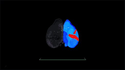
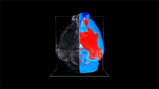
Whole Mouse Brain with c-Fos antibody labeling cleared using an organic solvent based procedure. The image was acquired on the Intelligent Imaging Innovations (3i) Cleared Tissue Lightsheet microscope. Acquired image in black and white. Registered regions from the Allen Brain Atlas in color. Automated cell detection and atlas registration performed on the NIH high performance cluster (Biowulf) using CATNIP code written by Dr. Snehashis Roy. Visualization created in Arivis Vision4D software.